45 microscope diagram with labels
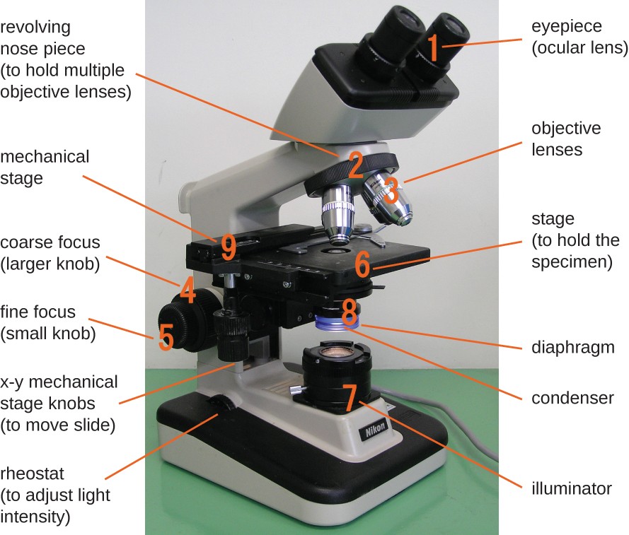
Label the microscope — Science Learning Hub Label the microscope Interactive Add to collection Use this interactive to identify and label the main parts of a microscope. Drag and drop the text labels onto the microscope diagram. eye piece lens diaphragm or iris coarse focus adjustment stage base fine focus adjustment light source high-power objective Download Exercise Tweet Label Microscope Diagram - EnchantedLearning.com diaphragm - an adjustable opening under the stage, allowing different amounts of light onto the stage. eyepiece - where you place your eye. fine focus adjustment - a knob that makes small adjustments to the focus (it is often smaller than the coarse focus knob). high-power objective - a large lens with high magnifying power.
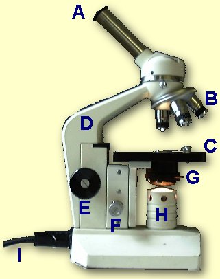
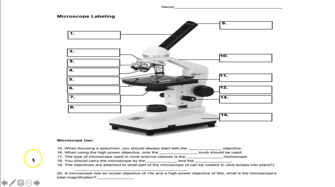
PDF Label parts of the Microscope: Answers Label parts of the Microscope: Answers Coarse Focus Fine Focus Eyepiece Arm Rack Stop Stage Clip . Created Date: 20150715115425Z ...

Microscope diagram with labels
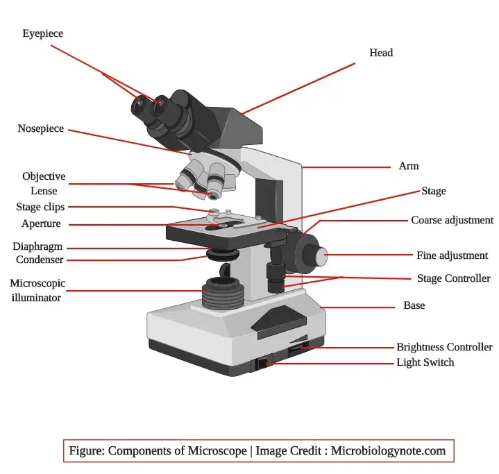
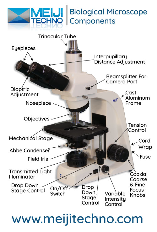
Compound Microscope Parts, Functions, and Labeled Diagram Eyepiece (ocular lens) with or without Pointer: The part that is looked through at the top of the compound microscope. Eyepieces typically have a magnification between 5x & 30x. Monocular or Binocular Head: Structural support that holds & connects the eyepieces to the objective lenses. Arm: Supports the microscope head and attaches it to the base. Label the microscope Diagram | Quizlet Diaphragm. Regulates the amount of light on the specimen. Light Source. Projects light upwards through the diaphragm, the specimen, and the lenses. Arm. supports the body tube. Stage. Supports the slide being viewed. Coarse Adjustment. Compound Microscope - Diagram (Parts labelled), Principle and Uses See: Labeled Diagram showing differences between compound and simple microscope parts Structural Components The three structural components include 1. Head This is the upper part of the microscope that houses the optical parts 2. Arm This part connects the head with the base and provides stability to the microscope.
Microscope diagram with labels. Sperm Under Microscope with Labeled Diagram - AnatomyLearner Sperm under microscope 400x labeled. I will show you the sperm under a microscope 400x with the labeled diagram. Here in the diagram, you will see some seminiferous tubules lined by the thick germinal epithelium. The picture shows the dark Type A and pale Type B spermatogonia located at the seminiferous tubules' basal part. Labeling the Parts of the Microscope | Microscope World Resources Labeling the Parts of the Microscope This activity has been designed for use in homes and schools. Each microscope layout (both blank and the version with answers) are available as PDF downloads. You can view a more in-depth review of each part of the microscope here. Download the Label the Parts of the Microscope PDF printable version here. Compound Microscope Parts - Labeled Diagram and their Functions What is a "compound microscope"? Labeled diagram of a compound microscope Major structural parts of a compound microscope Optical components of a compound microscope Eyepiece Eyepiece tube Objective lenses Nosepiece Specimen stage Coarse and fine focus knobs Rack stop Illuminator Condenser Abbe condenser Iris Diaphragm Condenser Focus Knob Summary Microscope Types (with labeled diagrams) and Functions Simple microscope labeled diagram Simple microscope functions It is used in industrial applications like: Watchmakers to assemble watches Cloth industry to count the number of threads or fibers in a cloth Jewelers to examine the finer parts of jewelry Miniature artists to examine and build their work Also used to inspect finer details on products
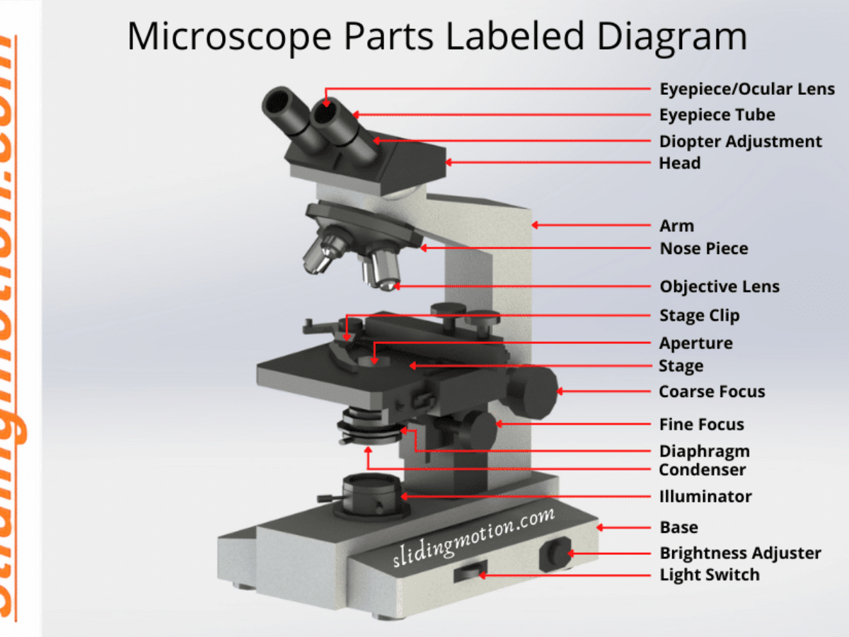
Microscope labeled diagram - SlideShare Microscope labeled diagram 1. The Microscope Image courtesy of: Microscopehelp.com Basic rules to using the microscope 1. You should always carry a microscope with two hands, one on the arm and the other under the base. 2. You should always start on the lowest power objective lens and should always leave the microscope on the low power lens ... Binocular Microscope Anatomy - Parts and Functions with a Labeled Diagram Ocular lens or eyepiece of the microscope, Diopter adjustment of the eyepiece All of these parts are identified in a light microscope labeled diagram. So, first, make sure you can identify all these parts from this labeled diagram. Parts of the compound microscope Microscope Parts, Function, & Labeled Diagram - slidingmotion Microscope Parts Labeled Diagram The principle of the Microscope gives you an exact reason to use it. It works on the 3 principles. Magnification Resolving Power Numerical Aperture. Parts of Microscope Head Base Arm Eyepiece Lens Eyepiece Tube Objective Lenses Nose Piece Adjustment Knobs Stage Aperture Microscopic Illuminator Condenser Lens Simple Microscope - Diagram (Parts labelled), Principle, Formula and Uses A simple microscope consists of Optical parts Mechanical parts Labeled Diagram of simple microscope parts Optical parts The optical parts of a simple microscope include Lens Mirror Eyepiece Lens A simple microscope uses biconvex lens to magnify the image of a specimen under focus.
Microscope Parts and Functions Body tube (Head): The body tube connects the eyepiece to the objective lenses. Arm: The arm connects the body tube to the base of the microscope. Coarse adjustment: Brings the specimen into general focus. Fine adjustment: Fine tunes the focus and increases the detail of the specimen. Nosepiece: A rotating turret that houses the objective lenses. PDF Parts of a Microscope Printables - Homeschool Creations Label the parts of the microscope. You can use the word bank below to fill in the blanks or cut and paste the words at the bottom. Microscope Created by Jolanthe @ HomeschoolCreations.net. Parts of a eyepiece arm stageclips nosepiece focusing knobs illuminator stage objective lenses Microscope, Microscope Parts, Labeled Diagram, and Functions The Microscopes parts divided into three different structural parts Head, Base, and Arms. Head/Body: It contain the optical parts in the upper part of the microscope. Arm: It supports the tube and connects it to the base. Base: The bottom of the microscope, used for support. Optical Components of Microscope A Study of the Microscope and its Functions With a Labeled Diagram ... A Study of the Microscope and its Functions With a Labeled Diagram To better understand the structure and function of a microscope, we need to take a look at the labeled microscope diagrams of the compound and electron microscope. These diagrams clearly explain the functioning of the microscopes along with their respective parts.
Draw a labelled diagram of a compound microscope.
Parts of a microscope with functions and labeled diagram - Microbe Notes Structural parts of a microscope and their functions Figure created with biorender.com Figure: Diagram of parts of a microscope There are three structural parts of the microscope i.e. head, base, and arm. Head - This is also known as the body. It carries the optical parts in the upper part of the microscope. Base - It acts as microscopes support.

draw a well label diagram of microscope - Brainly.in
The Parts of a Microscope (Labeled) Printable - TeacherVision The Parts of a Microscope (Labeled) Printable. Download. Add to Favorites. Share. This diagram labels and explains the function of each part of a microscope. Use this printable as a handout or transparency to help prepare students for working with laboratory equipment. Grade:
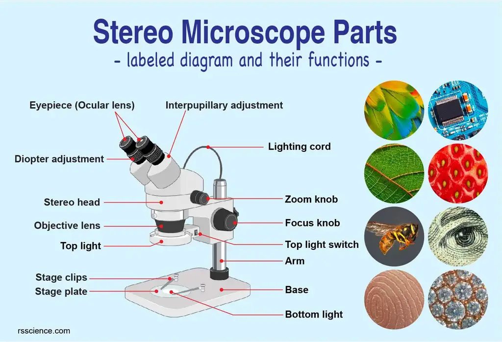
Parts of Stereo Microscope (Dissecting microscope) – labeled ...
Compound Microscope - Diagram (Parts labelled), Principle and Uses See: Labeled Diagram showing differences between compound and simple microscope parts Structural Components The three structural components include 1. Head This is the upper part of the microscope that houses the optical parts 2. Arm This part connects the head with the base and provides stability to the microscope.

Draw a neat labelled diagram of a compound microscope class ...
Label the microscope Diagram | Quizlet Diaphragm. Regulates the amount of light on the specimen. Light Source. Projects light upwards through the diaphragm, the specimen, and the lenses. Arm. supports the body tube. Stage. Supports the slide being viewed. Coarse Adjustment.
Label a microscope - Teaching resources
Compound Microscope Parts, Functions, and Labeled Diagram Eyepiece (ocular lens) with or without Pointer: The part that is looked through at the top of the compound microscope. Eyepieces typically have a magnification between 5x & 30x. Monocular or Binocular Head: Structural support that holds & connects the eyepieces to the objective lenses. Arm: Supports the microscope head and attaches it to the base.

Label the Microscope Diagram | Download Scientific Diagram

Parts of a Compound Microscope and Their Functions
Parts of a Microscope and Their Functions

Microscope With Labels clip art | Microscope parts ...

Simple Microscope Definition, Magnification, Parts And Uses

microscope drawing with label - Clip Art Library

Microscope Labeling Diagram | Quizlet

A labeled diagram of a microscope. MLT 101. :) | Medical lab ...

easy compound microscope diagram - Clip Art Library
Microscope with anatomy structure of Algae on white ...

How to draw Microscope diagram for beginners - step by step
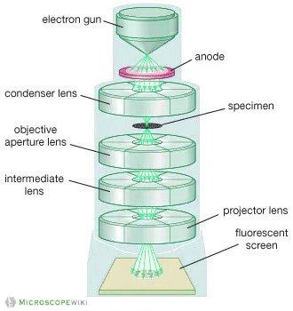
Electron Microscope Principle, Uses, Types and Images ...
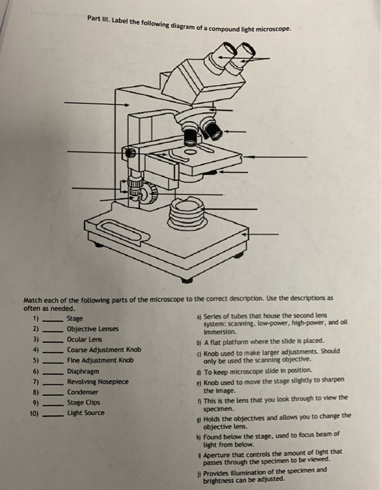
Solved Part III. Label the following diagram of a compound ...
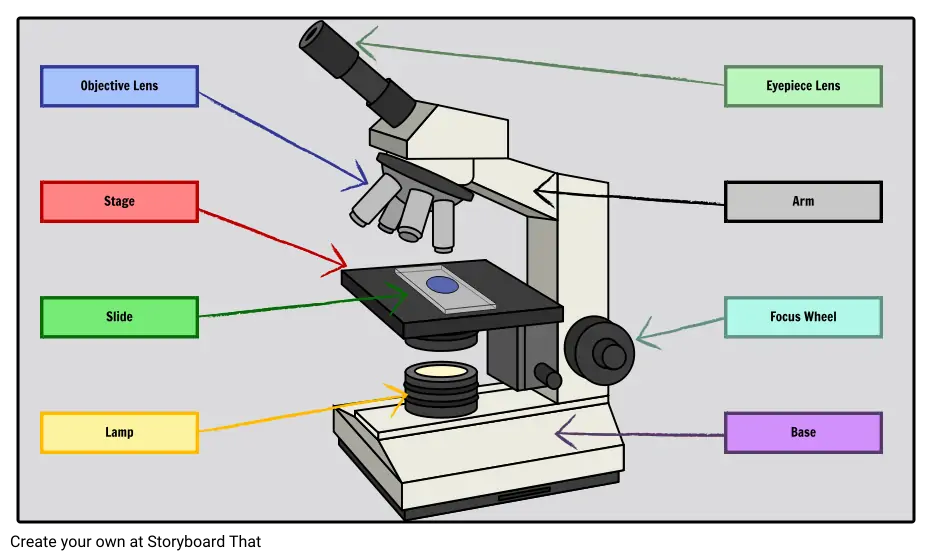
Labeled Microscope Storyboard by oliversmith
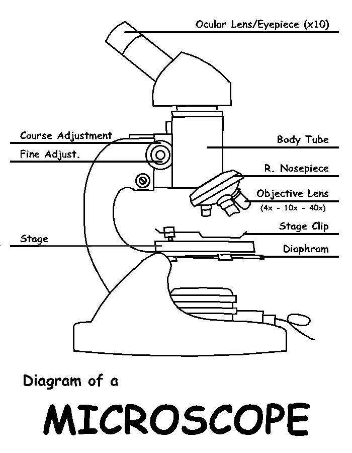
Diagram of a Microscope by ScienceDoodles on DeviantArt
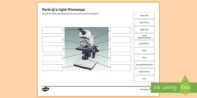
Parts of a Light Microscope Activity | Labeling Task
Microscope Terminology
Parts of Microscope, Function, Names & Labeled Diagram ...

Compound Microscope: Parts of Compound Microscope

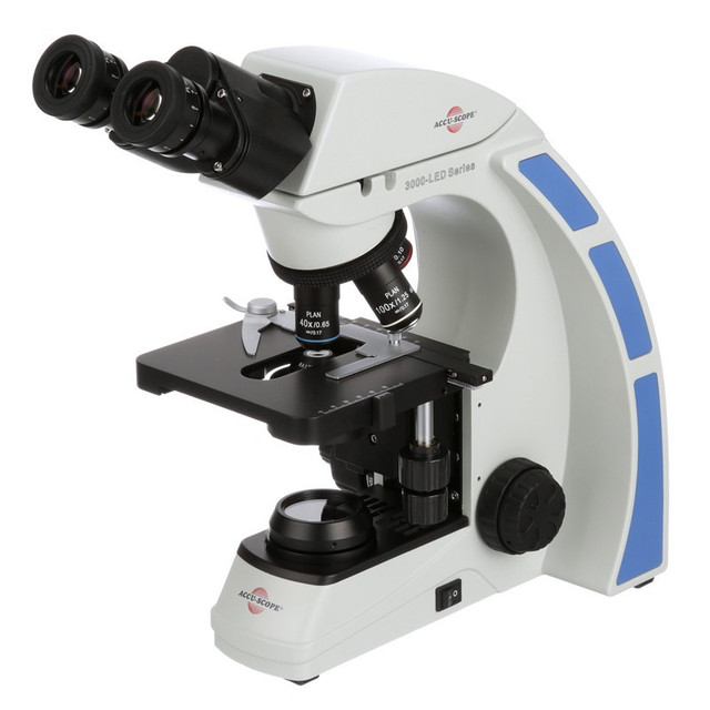
Produk Microscope | UD Berkah Abadi

Label Microscope Diagram - EnchantedLearning.com
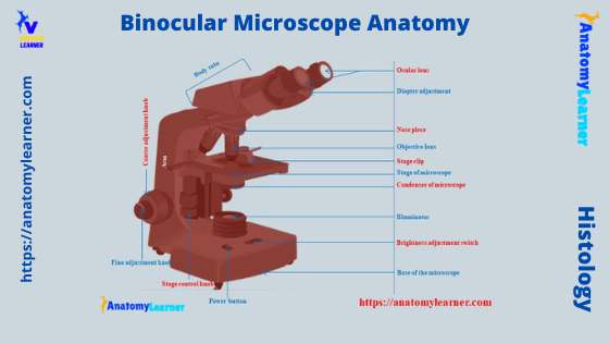
Binocular Microscope Anatomy - Parts and Functions with a ...

Microscope Diagram - Label Diagram | Quizlet

Compound Microscope- Definition, Labeled Diagram, Principle ...

label microscope diagram | Charts | Microscope, Anatomy bones ...
Junior cert Labelling Microscope - Labelled diagram

Diagram of a Compound Microscope

Living Environment Course

Microscope parts 3D learning for Android - APK Download

Compound Microscope Parts – Labeled Diagram and their ...

Microscope Labeling Activity - SMART Board Activity - Interactive Review

The Science Break - Labels for the light microscope for GCSE ...
Microscope
Compound Microscope Parts, Functions, and Labeled Diagram ...

File:Microscope diagram.png - Wikimedia Commons
Instruments of Microscopy | Microbiology | | Course Hero

Microscope, Microscope Parts, Labeled Diagram, and Functions
Microscope Terminology

Light Microscope- Definition, Principle, Types, Parts ...
Microscope Labeling


Komentar
Posting Komentar